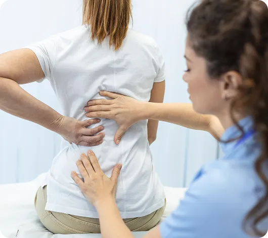
Spinal Stenosis Treatment
Relieve nerve pressure and restore mobility with minimally invasive care

What Is Spinal Stenosis?
Spinal stenosis occurs when the spaces within the spine become narrowed, putting pressure on the spinal cord and nerves. This narrowing can cause pain in the lower back and legs, especially while standing or walking, and may lead to balance issues, weakness, or coordination difficulties.
Also referred to as lumbar spinal stenosis, this condition can result from a variety of causes, including spinal injuries, thickened ligaments, herniated discs, or degenerative changes such as osteoarthritis. Its development is influenced by age, genetics, and lifestyle factors.
Early diagnosis is key to preventing progression and preserving mobility. Spinal stenosis is typically identified through a combination of physical exams and imaging studies.

Common Signs of Spinal Stenosis
Symptoms often develop gradually and may include:
Pain in the legs or lower back that worsens when standing or walking, and improves with rest
Tingling or numbness in the back, feet, or legs
Weakness in the legs or feet
Difficulty with balance or coordination
What Causes Spinal Stenosis?
Spinal stenosis can result from degenerative changes, congenital conditions, or trauma. Below are the most common causes:
Degenerative Changes (Spondylosis)
As we age, the spine naturally begins to wear down. This degeneration may lead to:
✔ Osteoarthritis and bone spurs: As cartilage deteriorates, bony growths can form, narrowing the space between joints.
✔ Disc degeneration: As discs lose height and hydration, they reduce the space for spinal nerves, leading to compression and discomfort.
✔ Facet joint hypertrophy: Enlargement of the joints in the spine can crowd the spinal canal.
✔ Herniated discs: When disc material pushes out of place, it can press on nerves.
✔ Thickened ligaments: Ligaments may become stiff and enlarged over time, further narrowing the spinal canal.
Congenital Factors and Trauma
Congenital factors can play a crucial role in the development of spinal stenosis. This disease can also be caused due to physical trauma:
✔ Spinal tumors: Some individuals may develop growths within or around the spinal cord due to genetic predisposition.
✔ Injury: Trauma from falls, workplace accidents, or car crashes can damage the spine and contribute to spinal canal narrowing or herniated discs.
How Is Spinal Stenosis Diagnosed?
In order to diagnose spinal stenosis, a health specialist must identify any congenital factors that could be contributing to the disease’s development. Determining genetic factors may lead to early diagnosis and prompt treatment, therefore, avoiding complications.
Then, a physical examination will be done for your lower back area, neck, and legs to identify any potential issues. If no anomalies are recognized with the physical inspection, your doctor will continue with imaging studies like X-rays, MRIs, and CT scans of your lower back area.
After examining your evaluations, your doctor will provide you with a diagnosis and treatment plan tailored to your specific needs.
Treatment Options for Spinal Stenosis
Treatment depends on the severity of the condition and how it affects your mobility and daily function. Options range from conservative therapies to minimally invasive or surgical procedures.
Therapies and Lifestyle Changes
Targeted physical therapy can increase mobility and reduce nerve pressure. Your care plan may include:
✔ Exercise: Low-impact activities like walking, swimming, or cycling help strengthen the back and improve flexibility.
✔ Weight management: Maintaining a healthy weight reduces spinal pressure and nerve irritation.
✔ Posture and ergonomics: Using ergonomic chairs and correcting sitting posture can ease stress on the lower back.
Lifestyle Changes
Some tips that could help alleviate the symptoms of degenerative disc disease include:
✔ Performing low-impact exercises to strengthen the muscles around the spine and add flexibility to the lumbar area
✔ Following a balanced diet and healthy lifestyle to manage weight and reduce lumbar stress
✔ Using ergonomic chairs and having good posture reduces pressure and pain in your spine
Medications and Injections
Over-the-counter and prescription medications can reduce spinal inflammation and nerve pressure. Your provider will tailor the dosage and type based on your symptoms.
Steroid injections, guided by imaging, are administered near the affected nerves to reduce inflammation and block pain signals. Injected into the epidural space, they offer longer-lasting relief than local anesthetics.
Injections
Unlike local anesthetics, injections offer longer-lasting relief for patients dealing with degenerative disc disease. Available options can include the following:
✔ Platelet-rich plasma (PRP) is derived from the patient’s own blood and contains multiple substances that promote tissue growth and regeneration.
✔ Stem cell therapy uses stem cells from multiple tissues of the patient’s own body and promotes tissue regeneration.
Pain Management Procedures
These procedures aim to disrupt pain signals and provide rapid relief, often used alongside therapy or medication:
✔ Radiofrequency ablation: Uses heat to deactivate specific pain-causing nerves.
✔ Nerve root blocks: Injections that block pain at its source by targeting spinal nerve roots.
✔ Facet joint injections: Administered directly into affected spinal joints to reduce inflammation.
✔ Pulsed radiofrequency therapy: Delivers high-frequency current to desensitize pain-generating nerves.
Neuromodulation
Neuromodulation is the medical technique of altering nerve activity using electrical stimulation. Its objective is to block the pain signals sent into the brain. These can help reduce the lumbar stress caused by spinal stenosis. Spinal cord stimulation, for example, is a treatment that uses electrical impulses to modify the nerve activity in the spine. As the nerve signals become blocked by this stimulation, the pain won’t be felt even though the symptom source remains.
Minimally Invasive Interventions
These outpatient procedures are designed to decompress nerves and restore function with minimal disruption:
✔ MILD® procedure (Minimally Invasive Lumbar Decompression): Uses imaging guidance to remove small amounts of bone or ligament that are compressing nerves.
✔ Vertiflex (interspinous spacers): Small implants placed between vertebrae to create space and relieve pressure on spinal nerves.
Surgical Treatments
Surgery may be considered when other treatments are ineffective:
✔ Decompressive laminectomy: Removes a portion of the vertebral arch to relieve spinal cord compression.
✔ Spinal fusion: Replaces a damaged disc with a bone graft to stabilize the spine.
✔ Interspinous process devices: Implants inserted between vertebrae to maintain space and prevent nerve compression.

Care That’s Close to Home
We offer care from two convenient clinic locations, making it easy to access expert medical support close to home.
Each facility is designed to provide a welcoming, safe, and efficient environment equipped with advanced technology and supported by a compassionate team dedicated to your well being.

Why Choose Spinal Diagnostics?
Patients choose Spinal Diagnostics for our comprehensive approach, accurate diagnostics, and compassionate care. We stay at the forefront of interventional procedures and are committed to improving your quality of life—without opioids or invasive surgeries.
Proven Medical Expertise
We bring years of clinical experience in pain management and interventional procedures.
Constant Innovation
We use the latest techniques and technology to ensure safe, effective treatment.
Compassionate Care
We listen, understand, and treat every patient with empathy and respect.
Personalized Plans
Each treatment is tailored to your condition, goals, and lifestyle.
Explore the treatments that bring real relief.
Learn how our non-invasive solutions and personalized plans can help you feel better, faster.
Explore the treatments that bring real relief.
Learn how our non-invasive solutions and personalized plans can help you feel better, faster.


